Home

opening Disgraceful deficit scanner sinus danger A good friend Authorization furniture

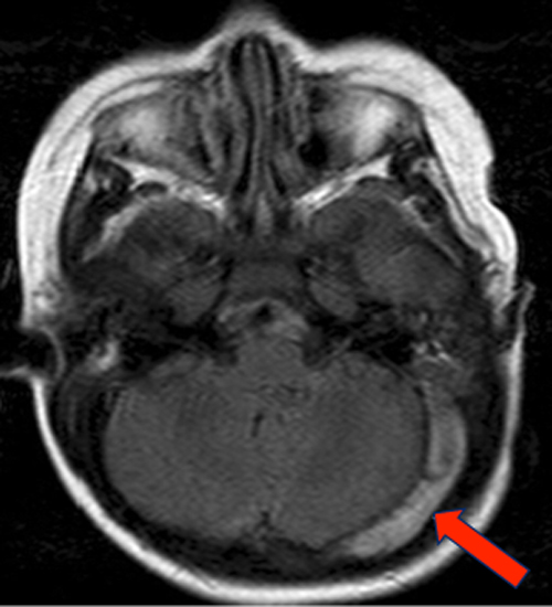
Clinical features of otogenic cerebral sinovenous thrombosis: Our  experience and review of literature - Abir - 2022 - Clinical Case Reports -  Wiley Online Library
Clinical features of otogenic cerebral sinovenous thrombosis: Our experience and review of literature - Abir - 2022 - Clinical Case Reports - Wiley Online Library

Orbital Infection Imaging: Practice Essentials, Computed Tomography,  Magnetic Resonance Imaging
Orbital Infection Imaging: Practice Essentials, Computed Tomography, Magnetic Resonance Imaging

Dental CT Scans – what you need to know

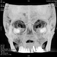
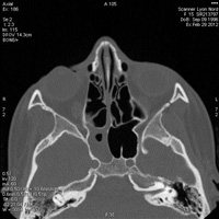
Scanner des Sinus de la Face à Lyon : Examen - Norimagerie

The type of the protrusion in the sphenoid sinus. NO protrusion in the... |  Download Scientific Diagram
The type of the protrusion in the sphenoid sinus. NO protrusion in the... | Download Scientific Diagram

Your child is having a CT scan with sedation | Great Ormond Street Hospital

Slow flow v thrombus - Questions and Answers ​in MRI

Relationship between sphenoid sinus volume and accessory septations: A 3D  assessment of risky anatomical variants for endoscopic surgery - Gibelli -  2020 - The Anatomical Record - Wiley Online Library
Relationship between sphenoid sinus volume and accessory septations: A 3D assessment of risky anatomical variants for endoscopic surgery - Gibelli - 2020 - The Anatomical Record - Wiley Online Library

Tomography | Free Full-Text | Patterns of Pneumatization of the Posterior  Nasal Roof
Tomography | Free Full-Text | Patterns of Pneumatization of the Posterior Nasal Roof

Sinus MRI scan
Sinus MRI scan

Scanner des Sinus de la Face à Lyon : Examen - Norimagerie

Third-, Fourth-, and Sixth-Nerve Lesions and the Cavernous Sinus |  SpringerLink
Third-, Fourth-, and Sixth-Nerve Lesions and the Cavernous Sinus | SpringerLink

Tomography | Free Full-Text | Patterns of Pneumatization of the Posterior  Nasal Roof
Tomography | Free Full-Text | Patterns of Pneumatization of the Posterior Nasal Roof

3-D Sinus Imaging - CT Scan of the Sinuses | NY Allergy & Sinus Centers

CT Scanning of the Paranasal Sinuses: Indications, Utilization, and  Radiation Risks - ENTtoday
CT Scanning of the Paranasal Sinuses: Indications, Utilization, and Radiation Risks - ENTtoday

Pre-FESS Imaging of Paranasal Sinuses and Nasal Cavity: Using  Multi-detector Computed Tomography (MDCT) in Understanding Normal Anatomy  and Anatomical Variations: Tips and Tricks | SpringerLink
Pre-FESS Imaging of Paranasal Sinuses and Nasal Cavity: Using Multi-detector Computed Tomography (MDCT) in Understanding Normal Anatomy and Anatomical Variations: Tips and Tricks | SpringerLink
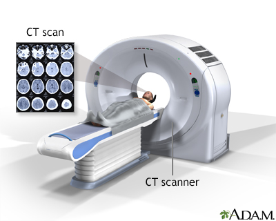
Head CT scan Information | Mount Sinai - New York

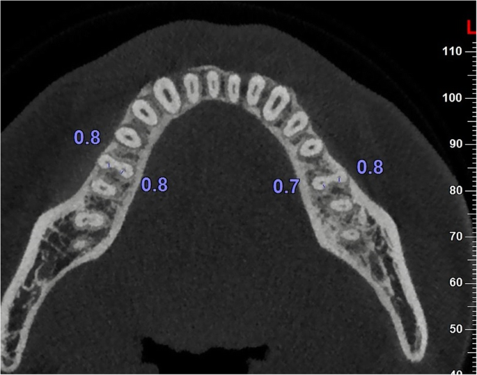
Root dentine thickness of danger zone in mesial roots of mandibular first  molars | BMC Oral Health | Full Text
Root dentine thickness of danger zone in mesial roots of mandibular first molars | BMC Oral Health | Full Text

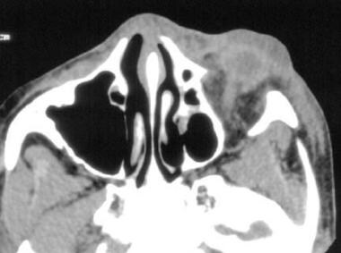
Brain MRI Scan | Tesla MRI Machine | Echelon Health

CT scan (with contrast) showing sphenoid sinus lesion with calcification. |  Download Scientific Diagram
CT scan (with contrast) showing sphenoid sinus lesion with calcification. | Download Scientific Diagram